Perinatal Research
TEAM:
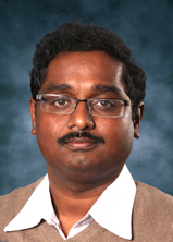 Jaladhar Neelavalli, PhD
Assistant Professor, Dept. Radiology and Biomedical Engineering
Dr. Neelavalli is interested in identifying hypoxic ischemic injury in the fetal brain using quantitative magnetic resonance imaging (MRI) methods. Measuring hypoxic-ischemic condition requires the ability to measure in-vivo blood oxygenation status as well as blood flow. Susceptibility Weighted Imaging (SWI) has been used clinically to evaluate such conditions in neonates and Dr. Neelavalli's group was the first to adapt this sequence to image the fetal brain. He has also applied the principles of MRI based susceptometry to measure venous blood oxygen saturation in the fetal brain, the first such measurement using MRI in human fetuses.
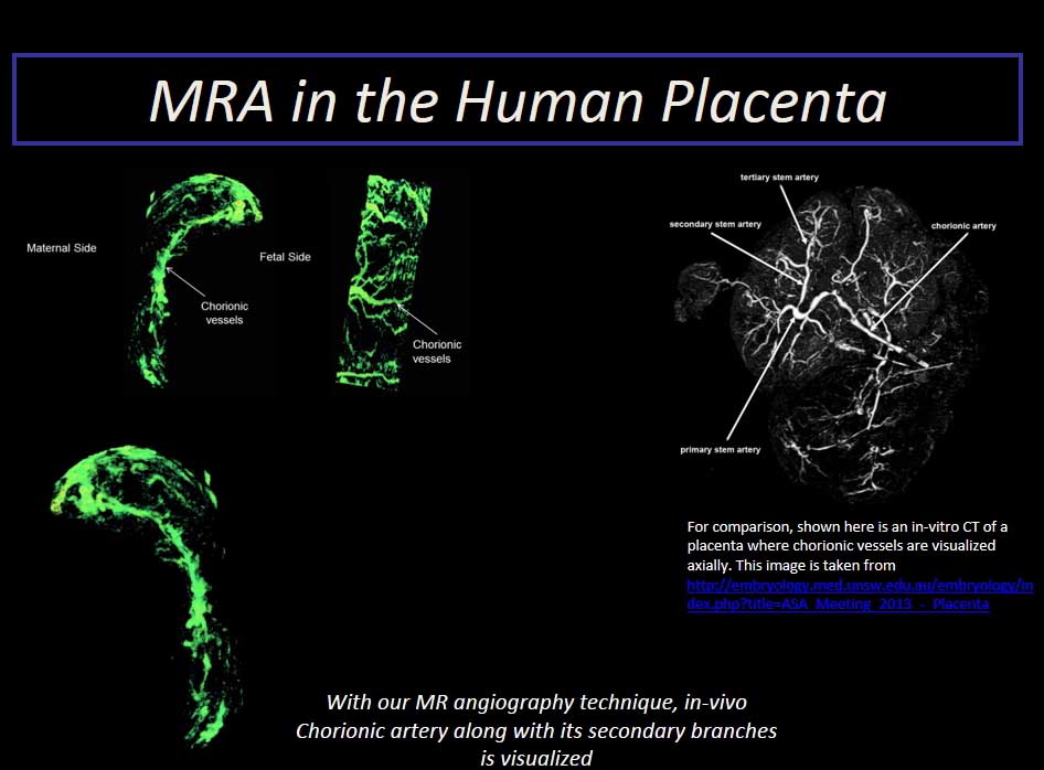
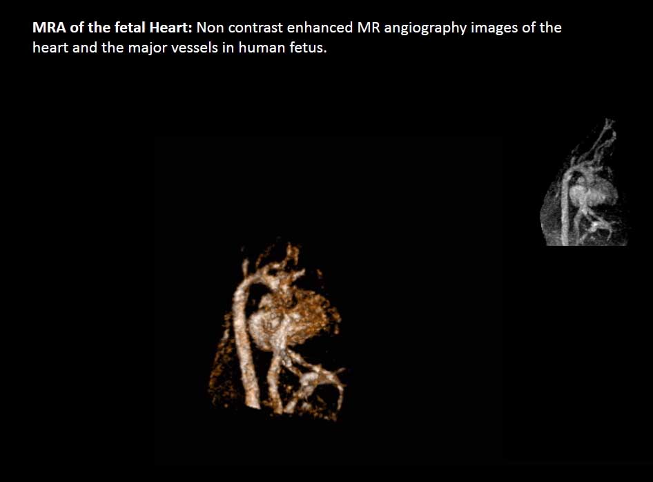
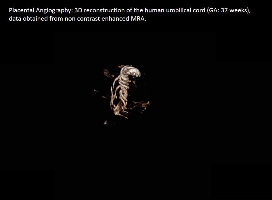
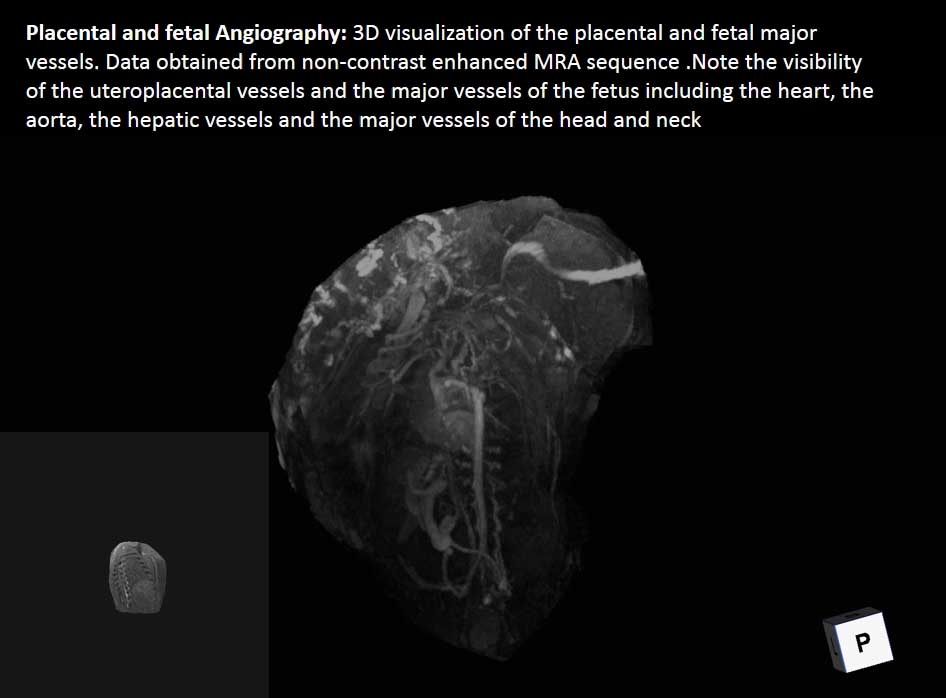
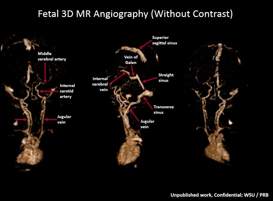
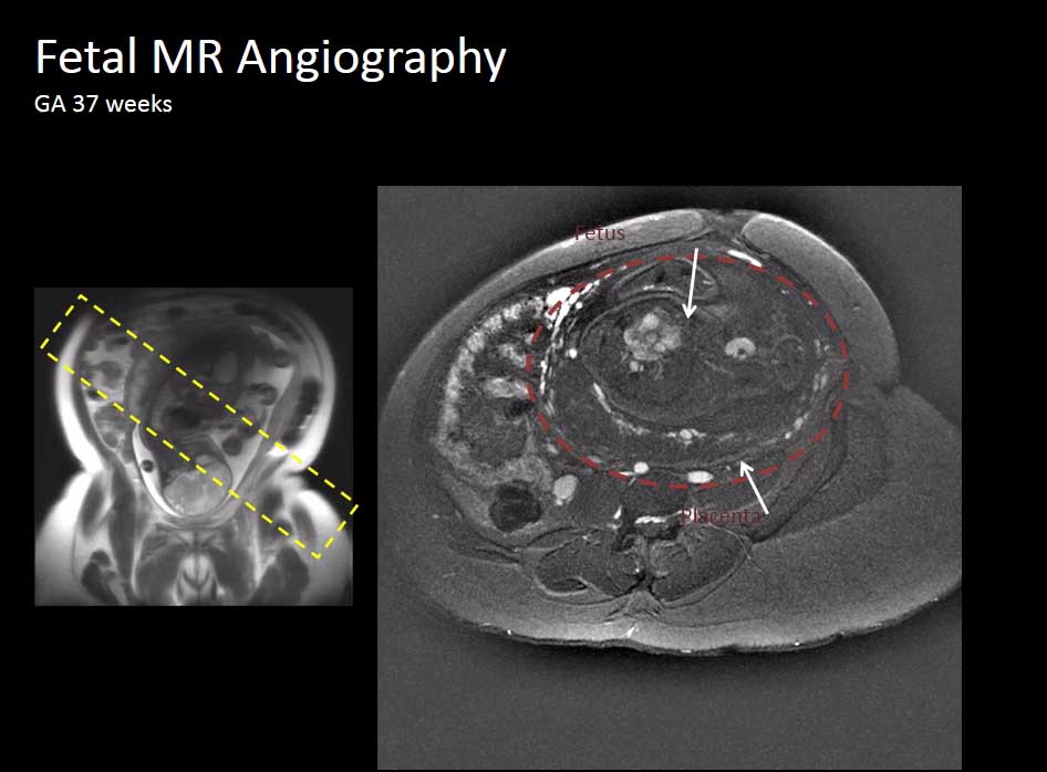
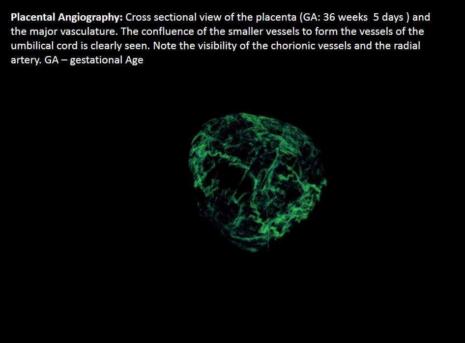
Angiographic imaging of feto-placental blood vessels is important in assessing vascular malformations. High resolution MR angiography (MRA) in-utero is possible clinically by appropriately optimizing the clinical MRI sequences. Faster and higher resolution angiography of in-utero structures is possible using advanced fast imaging methods. Feto-placental MRA is an important step in quantitative vascular imaging and developing fast fetal MRA techniques is one of the focuses of Dr. Neelavalli's group.
Using the imaging techniques usually applied in adult humans for imaging the human fetal brain is not straightforward, mainly due to fetal motion. As opposed to maternal respiratory motion which is periodic and relatively easy to address, fetal motion is random and unpredictable, and hence difficult to correct for. One of the ways to address this is to employ fast imaging methods that are resistant to motion. Dr. Neelavalli's focus is to develop and apply such methods. Fetal structures are small and hence require small voxel sizes (i.e., high resolution imaging) to sufficiently resolve different tissues. Imaging at high resolution with MRI is conventionally time consuming which on the other hand directly competes with the goal of fast fetal imaging. Dr. Neelavalli's group develops and applies MRI techniques that employ novel data acquisition techniques that facilitate faster imaging like non-Cartesian k-space sampling, segmented k-space acquisition and compressed sensing.
Dr. Neelavalli obtained his PhD in Biomedical Engineering at Wayne State University (WSU) with a focus on Magnetic Resonance Imaging. After completing his first postdoctoral fellowship at University of Oxford, UK, he joined WSU's MR Research Facility for further postdoctoral training. He subsequently became an Assistant Professorof Radiology with a secondary appointment in Biomedical Engineering. Dr. Neelavalli's earlier work focused on developing methods for quantifying tissue magnetic susceptibility non-invasively using MRI (quantitative susceptibility mapping). These methods are used for quantifying tissue iron content which have great application in studying various neurodegenerative diseases like Alzheimer's disease, Parkinson's disease, Multiple Sclerosis and even normal aging. He currently works in close collaboration with the faculty at the Perinatology Research Branch of NIH and the Children's Hospital of Michigan in the development and application of quantitative MRI methods for assessing fetal brain injury.
Google Scholar
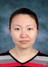 Feifei Qu, PhD
Postdoctoral Fellow
Dr. Qu joined our group in early 2016. She has an excellent background in MRI physics, experience in female pelvic and abdominal imaging, system programming skills in implementing fast imaging/reconstruction methods on MRI scanners and MR guided high intensity focused ultrasound therapy for uterine fibroids. She is currently focusing on further development of MRI techniques for fast flow quantification in fetal applications. HER interests lie in development of strategies for fast data collection and image reconstruction for structural imaging in general.
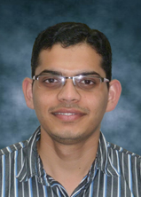 Uday Krishnamurthy, MS
Senior Ph.D. candidate, Department of Biomedical Engineering
Mr. Krishnamurthy's research is focused on developing MRI sequences for fetal imaging by (a) optimizing and (b) developing novel MR imaging sequences. He is working towards the development of a framework that allows for quantitative vascular imaging in the fetus based on (a) moving to higher field imaging while optimizing parameters for low energy deposition (b) developing faster sequences suited for fetal imaging and (c) development of non-Cartesian readout schemes to reduce scan time. Uday has addressed the safety concerns and shown the improvement in image quality when performing fetal imaging at 3.0T. In addition, he has developed a non-triggered phase contrast sequence to measure the blood flow non-invasively in fetus in-utero. He has been instrumental in demonstrating the role of time-of-flight MRA in fetal imaging and is currently working on accelerating the sequence and making it more robust.
Google Scholar
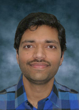 Brijesh Kumar Yadav, MS
PhD candidate, Department of Biomedical Engineering
Mr. Yadav has been a part of perinatal imaging research group and a member of the MRRF group at large for about three years now. His research efforts are towards making human-fetal MRI scans faster. This encompasses MR sequence design or optimization and development of data compression and penalty-based image reconstruction techniques for fetal-SWI scans. These efforts will lead to efficient quantification of some of the important human pregnancy-related physiological parameters such as fetal brain oxygenation. He was also involved in the animal study to evaluate changes in placental perfusion with advancing gestation in normal mice pregnancy using DCE-MRI.
Google Scholar
 Anabela Trifan, MBA
Study Coordinator
Ms. Trifan joined our group in mid 2015 and has been instrumental in managing patient enrollment and scanning support. She comes from an administrative and consulting background and has experience in training, management development, team collaboration, and communication. She is the central contact person for patients, our clinician collaborators and the lab members when it comes to patient recruitment and scanning. In addition to this, she also supports research grant preparation and submission.
VIDEOS:
Publications:
Neelavalli J*, Krishnamurthy U, Jella PK, Mody S, Yadav BK, Hendershot K, Hernandez-Andrade E, Yeo L, Cabrera M, Haacke EM, Hassan SS, Romero R. Magnetic Resonance Angiography of Fetal Vasculature at 3.0T. Eur Radiol. 2016 May 17. [Epub ahead of print]. PMID: 27189488.
Yadav BK, Neelavalli J*, Krishnamurthy U, Szalai G, Shen Y, Nayak NR, Chaiworapongsa T, Hernandez-Andrade E, Than NG, Haacke EM, Romero R. A longitudinal study of placental perfusion using dynamic contrast enhanced magnetic resonance imaging in murine pregnancy. Placenta. 2016 Jan 4. pii: S0143-4004(15)30115-6. doi: 10.1016/j.placenta.2015.12.019. [Epub ahead of print]. PMID: 26947613.
Krishnamurthy U, Szalai G, Shen Y, Xu Z, Brijesh K Yadav, Tarca A.L., Chaiworapongsa T, Hernandez-Andrade E, Than N.G, Haacke E.M, Romero R, Neelavalli J*. Longitudinal changes in placental MRI relaxation parameter in murine pregnancy: compartmental analysis. Gynecol Obstet Invest. 2016;81(3):193-201. doi: 10.1159/000431223. Epub 2015 Aug 26. PMID: 26336923.
Krishnamurthy U, Neelavalli J*, Mody S, Yeo L, Jella PK, Saleem S, Korzeniewski SJ, Cabrera MD, Ehterami S, Bahado-Singh RO, Katkuri Y, Haacke EM, Hernandez-Andrade E, Hassan SS, Romero R. MR imaging of the fetal brain at 1.5T and 3.0T field strengths: comparing specific absorption rate (SAR) and image quality. J Perinat Med. 2015 Mar 1;43(2):209-20.
Krishnamurthy U, Szalai G, Neelavalli J*, Shen Y, Chaiworapongsa T, Hernandez-Andrade E, Than N.G, Xu Z, Yeo L, Haacke E.M, Romero R. Quantitative T2 changes and susceptibility weighted magnetic resonance imaging in the murine pregnancy. Gynecol Obstet Invest. 2014;78(1):33-40.
Buch S, Liu S, Ye Y, Cheng YC, Neelavalli J, Haacke EM. Susceptibility mapping of air, bone, and calcium in the head. Magn Reson Med. 2015 Jun;73(6):2185-94. PMID: 25046134.
Neelavalli J*, Jella PK, Krishnamurthy U, Buch S, Haacke E.M., Yeo L, Mody S, Katkuri Y, Bahado-Singh RO, Hassan SS, Romero R, Thomason M. Measuring Venous Blood Oxygenation in Fetal Brain using Susceptibility Weighted Imaging. J Magn Reson Imag 2014 Apr;39(4):998-1006.
Liu S, Mok K, Neelavalli J*, Cheng YC, Tang J, Ye Y, Haacke E.M., Improved MR Venography Using Quantitative Susceptibility-Weighted Imaging. J Magn Reson Imaging. 2014 Sep;40(3):698-708. PMID: 24923249.
Neelavalli J*, Mody S, Yeo L, Korzenieski SJ, Saleem S, Katkuri Y, Jella PK*, Bahado-Singh RO, Hassan SS, Haacke EM, Romero R, Thomason M. Magnetic Resonance Venography Of The Fetal Brain Using Susceptibility Weighted Imaging. J Magn Reson Imag 2013. 2014 Oct;40(4):949-57.
2015
Best Scientific Presentation Award for Abstract # OC11.03: Quantitative blood flow estimation in the human fetus using non-triggered phase contrast magnetic resonance imaging. Krishnamurthy U, Feng W, Jella P, Yeo L, Hernandez-Andrade EA, Mody S, Cabrera MD, Haacke EM, Hassan SS, Romero R, Neelavalli J. 25th World Congress on Ultrasound in Obstetrics and Gynecology, Montreal, Canada
2015Summa Cum Laude Merit Award for Abstract #0945: Gestational Age Dependent Increase in Placental Perfusion Quantified Using MRI. Yadav BK , Neelavalli J, Krishnamurthy U, Shen Y, Szalai G, Wang B, Chaiworapongsa T, Hernandez-Andrade E, Than NG, Haacke EM, Hassan SS, Romero R. ISMRM 23rd Annual Meeting, Toronto, Ontario, Canada
2015Magna Cum Laude Merit Award for Abstract # 0638: Non-Contrast Magnetic Resonance Angiography of the Fetal Head and Neck Vessels. Krishnamurthy U, Neelavalli J, Jella PK, Hamtaei E, Mody S, Yadav BK, Hernandez-Andrade E, Yeo L, Cabrera MD, Haacke EM, Hassan SS, Romero R. ISMRM 23rd Annual Meeting, Toronto, Ontario, Canada
2016Best poster Award for Abstract: Imaging the Fetal Brain at 1.5T and 3.0T Field Strengths: Initial Experience with SAR and Image Quality Ultrasound Meets Magnetic Resonance. Krishnamurthy U, Neelavalli J, Mody S, Yeo L, Jella PK, Saleem S, Korzeniewski SJ, Cabrera MD, Ehterami S, Bahado-singh RO, Katkuri Y, Haacke EM, Hernandez-Andrade E, Hassan SS and, R. Romero at the European Society of Magnetic Resonance in Neuropediatrics Congress, Viena, Austria
Conference Abstracts:Krishnamurthy U, Feng W, Jella P, Yeo L, Hernandez-Andrade EA, Mody S, Cabrera MD, Haacke EM, Hassan SS, Romero R, Neelavalli J. Quantitative blood flow estimation in the human fetus using non-triggered phase contrast magnetic resonance imaging. Abstract # OC11.03, Proceedings of the 25th World Congress on Ultrasound in Obstetrics and Gynecology, Montreal, Canada, 2015. Best Scientific Presentation Award
Neelavalli J, Hernandez-Andrade EA, Yadav BK; Jella P; Krishnamurthy U; Yeo L; Mody S; Cabrera M; Haacke EM; Hassan SS; Romero R. Estimation of the cerebral metabolic rate of oxygen (CMRO2) in the human fetus. Abstract # OC11.08, Proceedings of the 25th World Congress on Ultrasound in Obstetrics and Gynecology, Montreal, Canada, 2015.
Neelavalli J, Sekar K, Hernandez-Andrade EA, Saker H, Krishnamurthy U, Yadav B, Haacke EM, Hassan S, Romero R. Evaluating synchronicity in binocular fetal eye movements using high resolution ultrasound. Abstract # OP09.03, Proceedings of the 25th World Congress on Ultrasound in Obstetrics and Gynecology, Montreal, Canada, 2015.
Neelavalli J, Krishnamurthy U, Jella P, Yadav B, Mody S, Hernandez-Andrade EA, Yeo L, Cabrera M, Haacke EM, Hassan S, Romero R. Non-contrast magnetic resonance angiography (MRA) in human pregnancy: visualization of the utero placental and fetal vasculature. Abstract # OP09.11, Proceedings of the 25th World Congress on Ultrasound in Obstetrics and Gynecology, Montreal, Canada, 2015.
Yadav BK , Neelavalli J, Krishnamurthy U, Shen Y, Szalai G, Wang B, Chaiworapongsa T, Hernandez-Andrade E, Than NG, Haacke EM, Hassan SS, Romero R. Gestational Age Dependent Increase in Placental Perfusion Quantified Using MRI. Proceedings of ISMRM 23rd Annual Meeting, Toronto, Ontario, Canada, poster# 0945, May 2015. Summa cum laude merit award.
Krishnamurthy U, Neelavalli J, Jella PK, Hamtaei E, Mody S, Yadav BK, Hernandez-Andrade E, Yeo L, Cabrera MD, Haacke EM, Hassan SS, Romero R. Non-Contrast Magnetic Resonance Angiography of the Fetal Head and Neck Vessels. Proceedings of ISMRM 23rd Annual Meeting, Toronto, Ontario, Canada, poster# 0638, May 2015. Magna cum laude merit award.
Neelavalli J, Krishnamurthy U, Jella PK, Mody S, Haacke EM, Roberto RR. In vivo Magnetic Resonance Angiography of the fetal vasculature. Society of Pediatric Radiology Annual Meeting – April 2015, Seattle, Washington.
Jella P, Krishnamurthy U, Neelavalli J, Mody S, Yeo L, Hernandez-Andrade E, Ehterami S, Cabrera M D, Korzeniewski S J, Haacke E.M., Hassan S, Romero R. Blood Oxygenation of Human Fetal Brain in the Second and Third Trimester. Proceedings of ISMRM-ESMRMB Joint Annual Meeting, Milano, Italy, poster# 0128, May 2014.
Katkuri Y, Buch S, Haacke EM, Latif Z, Neelavalli J, Xuan Y. Reducing the Background Field Variations Using the Geometry Information. AAPM 2014, Proceedings of 55th Annual Meeting & Exhibition, Indianapolis, Indiana. # WE-C-116-06.
Neelavalli J, Haacke E.M. Mody S, Yeo L., Saleem S. Katkuri Y, Jella P, Bahado-Singh R.O., Hassan S., Romero R, Thomason M. Magnetic Resonance Venography of the Fetal Brain Using Susceptibility Weighted Imaging (SWI). Proceedings of ISMRM 21st Annual Meeting, Salt Lake City, Utah, USA, poster# 0166.
Mody S, Yeo L, Neelavalli J, Thomason M, Hernandez-Andrade E, Hassan S. S, Romero R, Haacke E. M. (2013). Susceptibility weighted imaging of the fetal brain is superior to ultrasound and conventional MRI sequences in detecting intracranial hemorrhage. Proceedings of ESMRN 2013 conference titled Ultrasound meets Resonance Imaging in Vienna, Vienna. June 2013, Oral Presentation.
Feng W, Neelavalli J, Haacke EM, Hamtaei R, Katkuri Y, Xuan Y, Latif Z, Thomason ME. Measuring Flow in the Umbilical Cord Vessels Using Non-gated Phase Contrast MRI. RSNA Annual Meeting. Chicago, Illinois, USA, November 2012.
Krishnamurthy U, Neelavalli J, Mody S, Yeo L, Jella PK, Saleem S, Korzeniewski SJ, Cabrera MD, Ehterami S, Bahado-singh RO, Katkuri Y, Haacke EM, Hernandez-Andrade E, Hassan SS and, R. Romero. Imaging the Fetal Brain at 1.5T and 3.0T Field Strengths: Initial Experience with SAR and Image Quality Ultrasound Meets Magnetic Resonance, European Society of Magnetic Resonance in Neuropediatrics Congress, June 4-6 (2013), Vienna, Austria. Best poster Award